Turns a peripheral blood-smear image into reliability-scored, explainable evidence — reconstruction, cell detection and DSP pattern intelligence that pinpoint exactly what to verify. It screens and flags; a clinician confirms and decides.
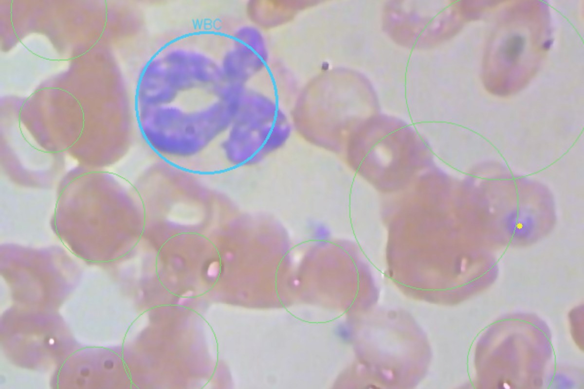
CellVision Blood reads a stained peripheral blood-smear image where a haematologist may not be in the room. It first reconstructs the image, then detects red cells, white cells and platelets, measures red-cell morphology, and runs an advanced DSP chain that pinpoints the most unusual cells and maps what it measures to recognised smear patterns — each shown with its evidence, a reliability score and the confirmatory test to order. It is deliberately not a diagnostic device: it supports a qualified reviewer and laboratory confirmation, and never replaces them.
Flat-field illumination correction, stain white-balance, denoise and acutance restore — the software analogue of a controlled scanner. Recovers cells lost to uneven lighting on cheap or field-condition optics.
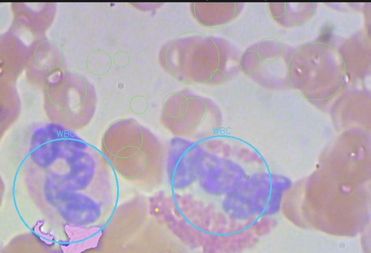
RBC counting by marker-controlled watershed (handles overlap), hue-based WBC-nucleus detection and platelet estimation — with per-cell size and central-pallor morphology.
Gabor-wavelet, GLCM/Haralick, LBP and radial-FFT chromatin descriptors per cell, scored by unsupervised novelty and clustered into subpopulations — then mapped to smear patterns to verify.
Measures how many more cells become detectable once the background is flattened — a concrete quality metric per image.
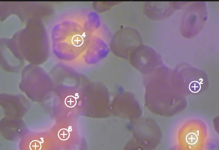
A rarity heatmap and ranked crosshairs pinpoint the cells that most deviate from the field — a triage “verify here” worklist.
Groups cells into morphological subpopulations so an unusual subgroup stands out at a glance.
Maps measured proxies to recognised patterns — anisocytosis, hypochromia, sickle-like, fragmentation, inclusions, leukocyte-rich — each with evidence and the confirmatory test.
A clean, explorable report: reconstructed image, detections, novelty map, population map, a proxy dashboard and the patterns-to-verify.
Per-image HTML report plus JSON for every measurement — nothing is a black box.
A reliability index accompanies every field and every pattern — a technical proxy for how much to trust a measurement, not calibrated statistical confidence. Soft or low-object images score low on purpose, urgent patterns are gated behind a reliability floor so the tool does not cry wolf, and every finding names the confirmatory test to order.
CellVision Blood screens and flags; it never outputs a diagnosis. A smear image can only reveal what is morphologically visible — it cannot measure blood chemistry, and its thresholds are engineered defaults, not clinical validation. A laboratory CBC and a qualified haematologist remain the source of truth.
Analysis runs locally — patient images never leave the workstation and there are no external API calls. The measurement core is deterministic and reproducible: the same image yields the same numbers, and every finding traces back to exactly where on the slide it came from.
CellVision Blood is a screening and decision-support prototype for the work around the diagnosis — especially where a specialist is not on site.
Reconstruction, detection and DSP intelligence, reliability-scored, explainable, offline-first and clinician-led.
Ground-truth datasets (starting with malaria, where public labels exist), calibrated confidence and measured precision/recall.
Scale calibration for real densities, a quality system, clinical studies and regulatory clearance before any diagnostic claim.
Screening programs, point-of-care clinicians, laboratory triage and teaching teams — a qualified reviewer and lab confirmation always stay in control.
The DSP novelty map with pinpointed cells to verify, alongside reconstructed detection views.

Talk to our team about a tailored walkthrough for your environment.
Talk to SalesResearch prototype. Every finding is a reliability-scored measurement, not a diagnosis; a qualified clinician and laboratory confirmation retain full interpretive responsibility. CellVision Blood is not a medical device and is not intended for clinical diagnosis. A smear image cannot assess blood chemistry (glucose/HbA1c, lipids, kidney/liver panels, hormones) or a “biological age”.
Built to fit inside the engineering, safety and compliance frameworks your organization already operates under.
Deployable inside an ISMS — data stays in your boundary, with access control and audit trails.
Supports condition- and risk-based asset decisions with defensible evidence.
Read-only, on-prem or private-cloud options keep sensitive data under your control.
Designed to support and align with the frameworks above. Alignment describes how the platform fits your program; it is not a claim of independent certification unless separately stated.
Cellvision Blood is designed to deliver value on day one for a small team, and to scale to a governed, multi-site enterprise deployment on the same platform.
Illustrative walkthroughs of how Cellvision Blood is used and the value it creates. Your figures are set on your own data during a proof-of-value engagement.
A scoped engagement on your own data shows the results before any wide rollout — measured, not promised.
Start with one team or site, then expand across the organization on the same platform — no re-buy, no re-build.
Every output is explainable and audit-ready, so risk and compliance teams can stand behind it.
A scoped engagement shows the results measured on your own operation before any wide rollout — then scale across the organization on the same platform.
Book a proof of value
